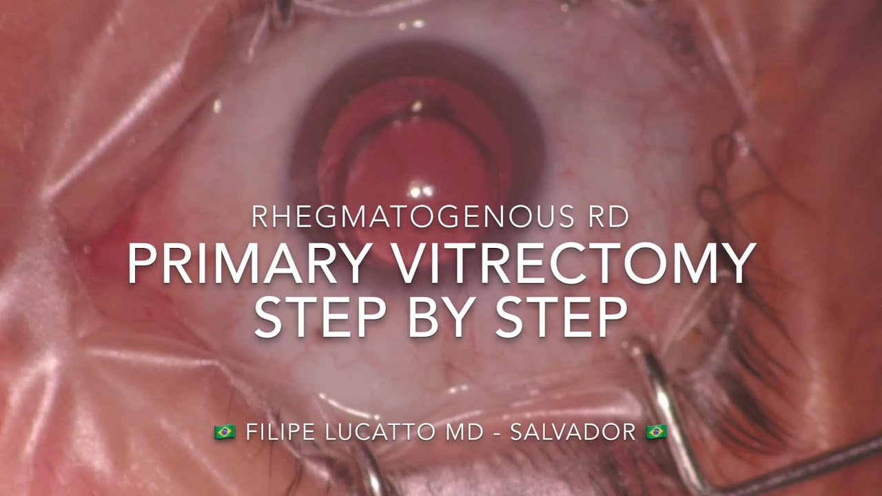
In this video we discuss the surgical steps in primary PPV for Rhegmatogenous retinal detachment
At first, a 23-gauge trocar is placed inferotemporal in a beveled uniplanar incision to insert the infusion line. Then, ensure the infusion line is fluid-filled, interrupt the flux and after connecting the infusion and check the correct position the infusion can be turned on. A second trocar is placed to connect a Chandelier light. In cases of hypotony or thicker sclera, a biplanar incision can be prefered. To place the remaining trocars, you can use bimanual technique, so you hold the globe with a hand while making the contralateral incision. A wide field non-contact view system was placed, and the focus adjusted to start the vitrectomy.
In the beginning of the surgery, before core vitrectomy, don't forget to remove the anterior vitreous behind the IOL and near the trocars. In this case, the preoperative US scans showed a total PVD but, even so, Triamcinolone was used in the surgery to ensure that areas of vitreoschisis are nor present. To shave the vitreous base, it’s important to use high cut rates and low vacuum to avoid iatrogenic breaks. A great advantage of using Chandelier light is to be able to indent to yourself and enable using higher vacuum near the depressed area. Patient had only small breaks in the inferior periphery and so it’s important to mark the breaks with a cautery before reattaching the retina.
PFCL was injected in a single bubble, to stabilize the posterior retina and allow a safer shaving of the vitreous base. During injection, It’s important to tilt the eye away from the breaks so the subretinal fluid will be slowly pushed through it.
In this case a Dual bore MedOne cannula was used for a more controlled injection, avoiding complications related with the jet stream and fish-eggs bubbles. After completing the shaving, the eye was filled up with PFLC and endolaser made inferiorly around the breaks.
During FAX, a soft tip cannula should be placed over the break, and all the fluid between the air bubble (above) and the perfluor bubble (under) needs to be aspirated first. Only when you have left just the heavy liquid and air, are you allowed to go posterior and aspirate the rest of PFCL near the optic disc. Sometimes in pseudophakic eyes the visualization can be worse during FAX due to fogging of the posterior face of the IOL. You can use a soft tip cannula to clean up the condensation of the IOL.
In this case remaining trapped subretinal fluid flowed toward the posterior pole and detached again the retina inferiorly. After local diathermy, a retinotomy was made to drain the subretinal fluid.
At the end of the surgery, 20% SF6 gas was injected as a tamponade.
Video:
Filipe Lucatto MD
Salvador - Brazil
Edition:
Filipe Lucatto MD
Juliana Prazeres MD
Salvador - Brazil
Neste vídeo, discutimos as etapas cirúrgicas da vitrectomia via pars plana para descolamento de retina regmatogênico.
Inicialmente, um trocater de 23-G é colocado TI em uma incisão uniplanar inclinada para inserir a infusão. Em seguida, certifique-se de que a linha de infusão está cheia de líquido, interrompa o fluxo, e após conecta-la, verifique a posição correta antes de liga-la. Um segundo trocater é colocado para conectar a Chandelier. Em casos de hipotonia ou esclera mais espessa, uma incisão biplanar pode ser preferida. Para colocar os trocateres restantes, você pode usar a técnica bimanual, de modo que segure o globo com a mão enquanto faz a incisão contralateral.
No início da cirurgia, antes da vitrectomia do core, não se esqueça de remover o vítreo anterior atrás da LIO e próximo aos trocateres.
Para o shaving da base vítrea, é importante usar altas taxas de corte e vácuo baixo, para evitar roturas. Usando a Chandelier, é possível indentar para si mesmo e permitir o uso de vácuo mais alto perto da área deprimida. É importante marcáaroturas pequenas com um cautério antes de reaplicar a retina. O PFCL foi injetado em uma única bolha, para estabilizar a retina e permitir shaving mais seguro. Durante a injeção, inclline o olho para longe das roturas para que o fluido sub seja empurrado lentamente através dele.
Neste caso, uma cânula Dual bore para evitar complicações relacionadas com a corrente de jato e fish-eggs. Após a finalização do shaving, o olho foi preenchido com PFLC e endolaser realizado.
Durante o FAX, uma soft tip deve ser colocada sobre a rotura, e todo o fluido entre o ar e o PFCL precisa ser aspirado primeiro. Somente depois de deixar apenas o PFCL e o ar, você pode aspirar o restante do PFCL próximo ao disco óptico. Em pseudofácicos, a visualização pode ser pior durante o FAX devido ao embaçamento da face posterior da LIO. Uma soft tip pode ser usada para limpar a condensação.
Nesse caso, o líquido sub fluiu em direção ao pólo posterior e a retina inferior descolou novamente. Após diatermia local, foi realizada retinotomia para drenagem do líquido sub.
Gás SF6 a 20% foi usado com tamponante.


![[Top Secret History] - Нарукавные щиты солдат Третьего Рейха (Эту награду первой срывали с пленных.)](http://i.ytimg.com/vi/RhwF3XkOZr8/mqdefault.jpg)


![[Top Secret History] - Нарукавные щиты солдат Третьего Рейха (Эту награду первой срывали с пленных.)](https://i.ytimg.com/vi/RhwF3XkOZr8/mqdefault.jpg)