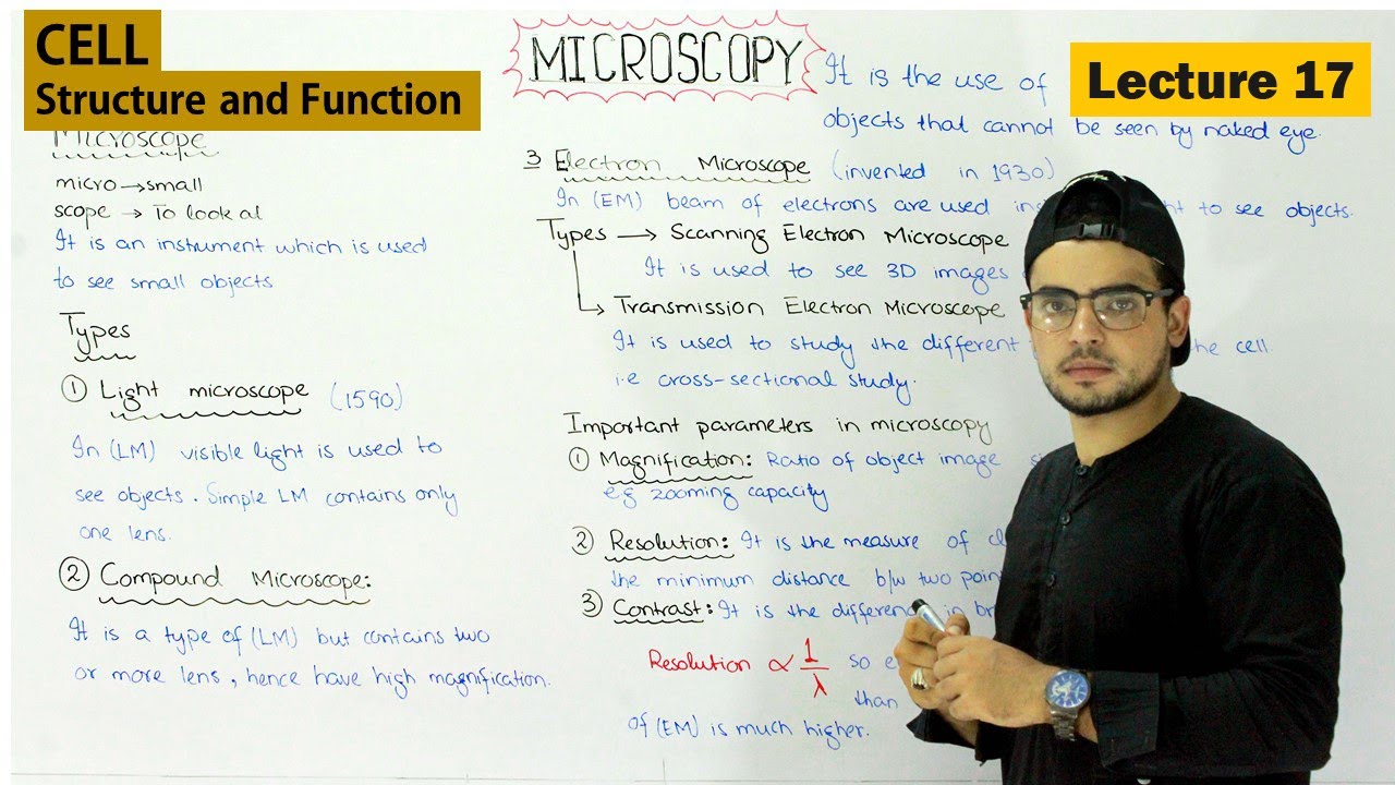
Microscopes were invented in 1590, In a light microscope (LM), visible light is passed through the specimen and then through glass lenses. The lenses refract (bend) the light in such a way that the image of the specimen is magnified as it is projected into the eye or into a camera.
In Compound microscope which is a type of light microscope two or more lenses are used for greater magnification. Three important parameters in microscopy are magnification, resolution, and contrast.
Magnification is the ratio of an object’s image size to its real size. Resolution is a measure of the clarity of the image; it is the minimum distance two points can be separated and still be distinguished as separate points. The third parameter, contrast, is the difference in brightness between the light and dark areas of an image. The electron microscope (EM) focuses a beam of electrons through the specimen or onto its surface.
Resolution is inversely related to the wavelength of the light (or electrons) a microscope uses for imaging, and electron beams have much shorter wavelengths than visible light. The scanning electron microscope (SEM) is especially useful for detailed study of the topography of a specimen. The transmission electron microscope (TEM) is used to study the internal structure of cells