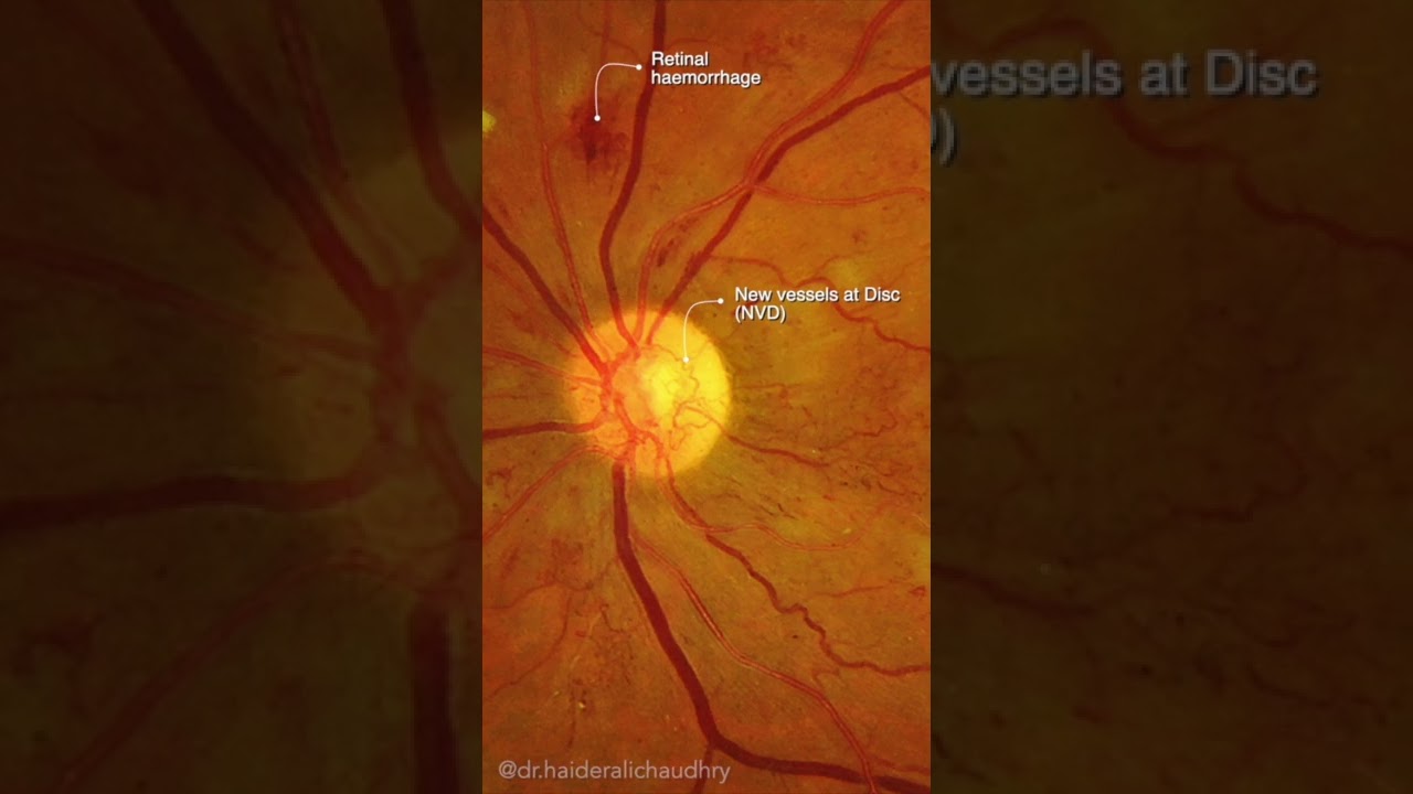
New vessels on disc (NVD) - defined as new vessels at or within 1 disc diameter of the disc, begins as fine loops or networks of vessels lying on the surface of the disc or bridging across the physiologic cup. They are usually easily identified once established, but in their earliest stages
they may be overlooked, especially with the low magnification of binocular indirect ophthalmoscopy. They also may be difficult to distinguish from normal vessels in nonstereoscopic photographs or with monocular direct ophthalmoscopy. The most satisfactory examination methods are those that provide a magnified stereoscopic view, using either biomicroscopy with contact or precorneal lens. Following points may come in handy in identification of subtle NVD
- their caliber is commonly one-eighth to one-quarter that of a major retinal vein at the disc margin.
- New vessel networks may also be irregular in shape, without a distinct radial pattern.
- New vessel patches often lie over retinal veins and appear to drain into them.
- New vessels have a unique capability of crossing both arterioles and veins in the underlying retina.
- Early in their evolution, new vessels appear bare, but later, delicate white fibrous tissue usually becomes visible adjacent to them.
- New vessels can readily be identified using fluorescein angiography during which they will leak profusely, unlike normal physiologic vasculature.
#shorts


![[4K] Middle East AI Lookbook-Arabian- Ethereal Pine Forest](http://i.ytimg.com/vi/r8SbMYdk3n8/mqdefault.jpg)