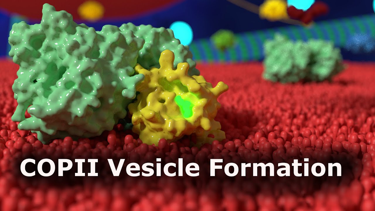
This animation shows the formation of a COPII vesicle and its transport to the Golgi apparatus.
Interesting talks about this topic:
Randy Schekman (Berkeley) Part 1: Studying Protein Secretion in Yeast (iBiology)
[ Ссылка ]
Randy Schekman (Berkeley) Part 2: Biochemical Reconstitution of Transport Vesicle Budding (iBiology)
[ Ссылка ]
Randy Schekman (Berkeley) Part 3: Human Diseases of Vesicle Budding (iBiology)
[ Ссылка ]
The Golgi Apparatus and ER to Golgi (Anterograde) Transport Part 1 - 4
[ Ссылка ]
Further readings:
COPII: a membrane coat formed by Sec proteins that drive vesicle budding from the endoplasmic reticulum. (Barlowe et. al., 1994)
COPII and COPI Traffic at the ER-Golgi Interface. (Szul and Szrul, 2011)
Assembly, organization, and function of the COPII coat. (Hughes and Stephens, 2007)
COPII and the regulation of protein sorting in mammals. (Zanetti et. al., 2012)
Music: The 126ers - Secret Conversations
This Video can be saved, shared or embedded for educational and non-commercial purpose.
Ribosome Studio is an animation studio based in Switzerland. We produce high-quality scientific animations, medical animations, and mechanism of action movies. For more information go to [ Ссылка ]
COPII Vesicle Formation
Теги
COPIIVesicleGolgi ApparatusTransport ProteinProtein TrafficingProtein TransportCell Biology (Field Of Study)Scientific AnimationScienceEndoplasmic Reticulum (Gene Group)KinesinMotor ProteinErgicendoplasmic reticulum-golgi intermediate compartmentCOPII vesicle formation animationCOP2vesicle transportcopII coated vesiclesVesicle buddingAnterograde TransportProtein transportmedical animationbiomedical animationhttp://www.ribosomestudio.com