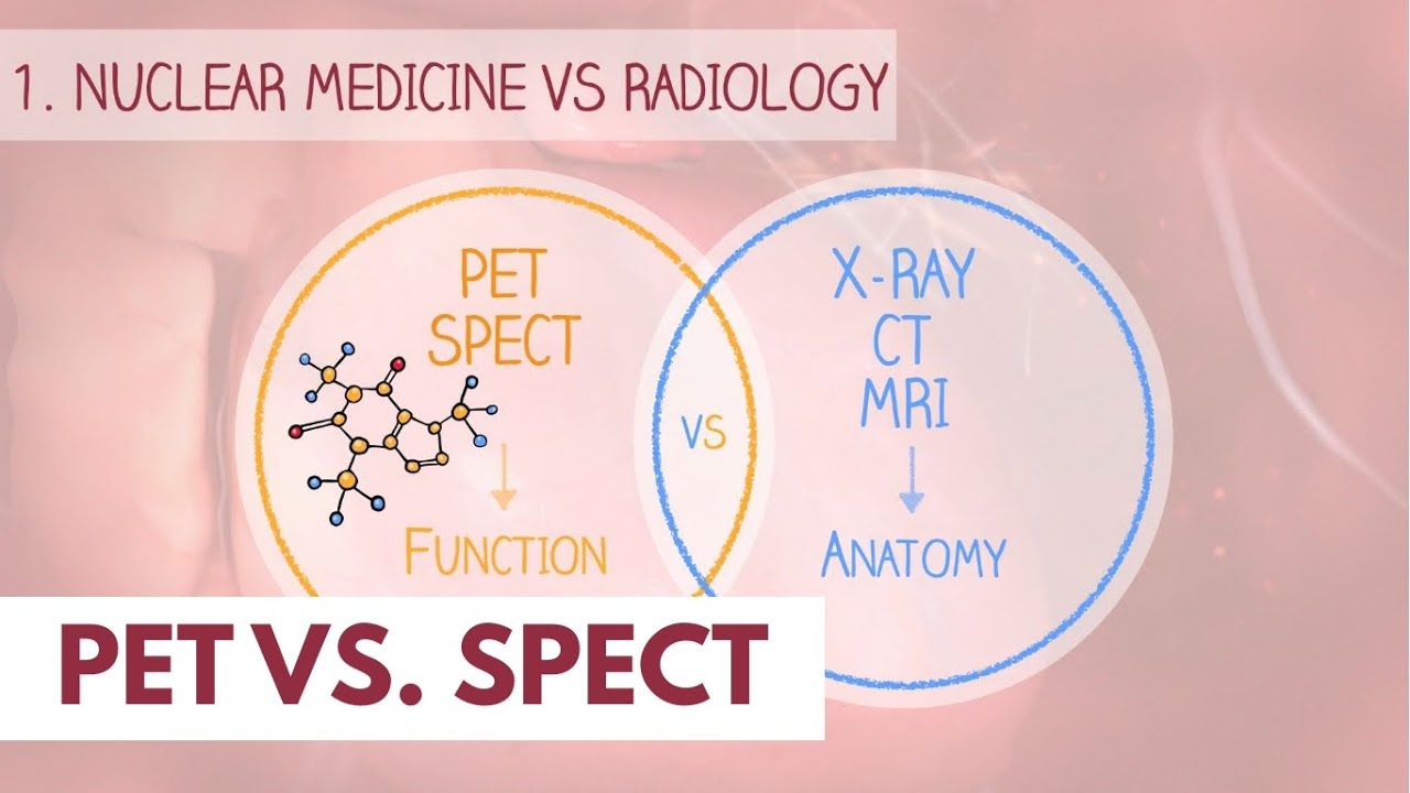
This video explains PET and SPECT, two primary imaging techniques in nuclear medicine that use small amounts of radioactive tracers to create detailed pictures of our bodies. Unlike X-rays, CT, or MRI scans that show the body's structure, PET and SPECT reveal how organs function, allowing for the detection of early biochemical changes and aiding in early disease diagnosis, particularly in oncology. The video covers the principles of PET, which uses positron-emitting radioisotopes like fluorine-18, and SPECT, which uses gamma-emitting radioisotopes like technetium, and how these radioisotopes are attached to tracers to target specific body processes. You'll learn how these radiotracers work inside the body to emit gamma rays, which are then captured by cameras to create detailed functional images.
LinkedIn: [ Ссылка ]
Instagram: [ Ссылка ]
Website: Coming soon! :-)
Timecodes:
0:00 Nuclear medicine vs. Radiology
0:28 Role of SPECT/PET
0:48 PET
1:12 SPECT
1:20 Radiotracers
1:38 Fluorodeoxyglucose
2:10 Technetium 99m-methyl Diphosphonate
2:35 Summary Tracers
2:46 PET - Image Creation (Annihilation)
3:45 SPECT - Image Creation
4:00 Next video
PET vs. SPECT scan | Dr. Paulien Moyaert
Теги
PETSPECTnuclear medicineimaging techniquesradioactive tracerspositron emission tomographysingle-photon emission tomographyorgan functionbiochemical processesearly disease detectiononcologycancer diagnosisfluorine-18technetiumradiotracersfluorodeoxyglucoseFDGmethylene diphosphonatebone scansgamma raysmedical imagingPET scannerSPECT scannerpositrongamma-emitting radioisotopesnuclear medicine playlistimagingImaging techniquesradiology