Audience: Radiology Residents
Learning Objectives:
Describe the difference between the forms of Doppler Imaging
Pulse wave Doppler (spectral wave / duplex)
Color Doppler
Power Doppler
M-mode imaging (not Doppler)
Summary:
Doppler US detects frequency shifts due to motion
Pulse wave (spectral) Doppler
Allows analysis of the velocity overtime
Small field of view (gate or sample volume)
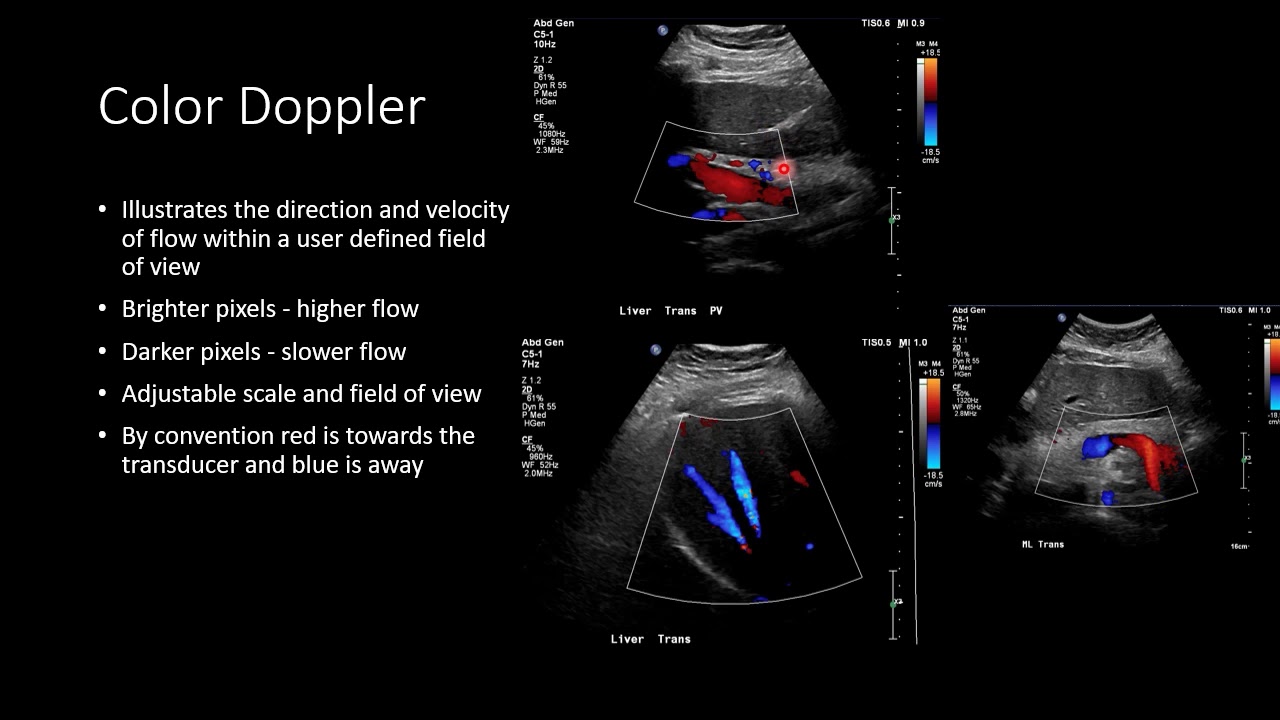
Color Doppler
Direction and velocity information encoded in 2 colors
Wide field of view
Power Doppler
Non-directional representation of flow
Higher sensitivity for slow flow
M-Mode – Not Doppler
Low power method for detecting rapid motion
1st trimester fetal heart rate
Cardiac valvular motion
References:
Hertzberg, Barbara S., and William D. Middleton. Ultrasound: the requisites. Elsevier Health Sciences, 2015.
Hangiandreou, Nicholas J. "AAPM/RSNA physics tutorial for residents: topics in US: B-mode US: basic concepts and new technology." Radiographics 23.4 (2003): 1019-1033.
Boote, Evan J. "AAPM/RSNA physics tutorial for residents: topics in US: Doppler US techniques: concepts of blood flow detection and flow dynamics." Radiographics 23.5 (2003): 1315-1327.


![This - That [Short Quiz]](https://i.ytimg.com/vi/2cvzG__o3RY/mqdefault.jpg)