📌 𝐅𝐨𝐥𝐥𝐨𝐰 𝐨𝐧 𝐈𝐧𝐬𝐭𝐚𝐠𝐫𝐚𝐦:- [ Ссылка ]
📌𝗝𝗼𝗶𝗻 𝗢𝘂𝗿 𝗧𝗲𝗹𝗲𝗴𝗿𝗮𝗺 𝗖𝗵𝗮𝗻𝗻𝗲𝗹 𝗛𝗲𝗿𝗲:- [ Ссылка ]
📌𝗦𝘂𝗯𝘀𝗰𝗿𝗶𝗯𝗲 𝗧𝗼 𝗠𝘆 𝗠𝗮𝗶𝗹𝗶𝗻𝗴 𝗟𝗶𝘀𝘁:- [ Ссылка ]
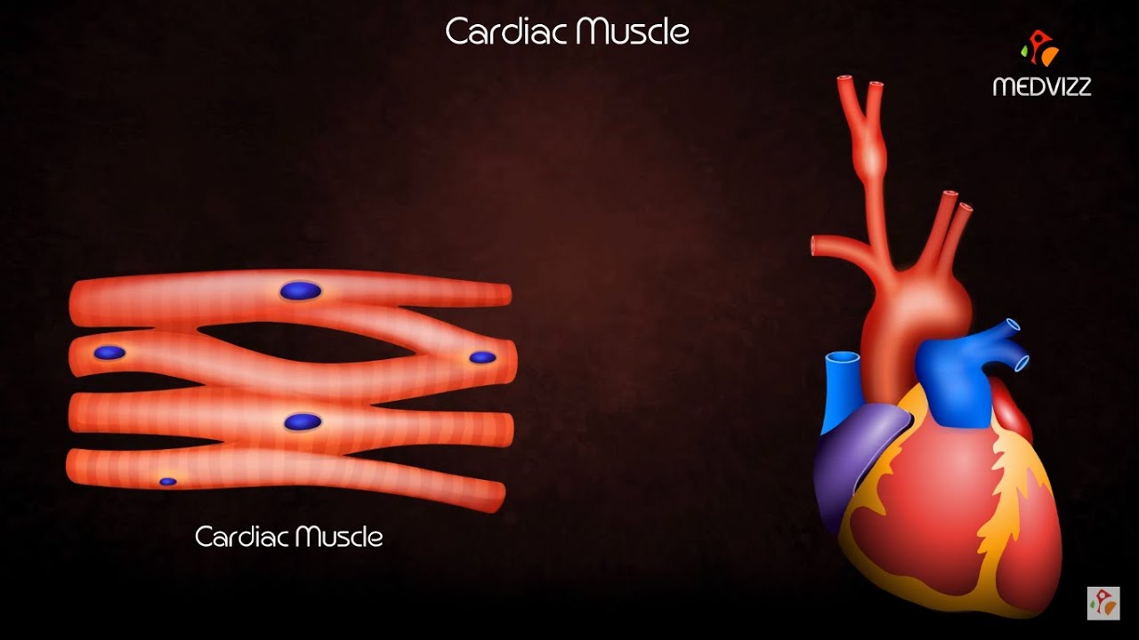
Cardiac Muscle Physiology - Usmle step 1 Quick review
It is very easy to overlook and take for granted a particular structure that is not readily visible in the human body. One such example is the muscles. It is very easy to observe skeletal muscle tissue, especially if you exercise physically.
However, smooth and cardiac muscle tissues are not so obvious compared to well-developed triceps or deltoids. However, you might guess that they are equally significant.
This article will start by describing the general classification of muscle tissue. After that, it will focus on the characteristics, components, and briefly on the contraction of cardiac muscle tissue.
Cardiomyocytes
--------------------------
Cardiomyocytes, also known as cardiac muscle cells, usually contain one elongated nucleus that lies in the center, which is a distinguishing feature from skeletal muscle. By examining the ultrastructure, it becomes apparent that the myofibrils separate as they approach the nucleus, pass around it, and re-assemble in their original pattern on the other side. You can visualize the arrangement by imagining two cones that are joined at their vertices, which represent the nucleus. In fact, cell organelles are also concentrated in this cytoplasmic region around the nucleus. These include mitochondria, Golgi apparatus, lipofuscin filled granules, and glycogen. Lipofuscin is a red-brown pigment, often called the wear and-tear-pigment, which gradually accumulates inside cardiac tissue with age. It is the remnant of lysosomal cell contents. The cytoplasm of cardiomyocytes, called sarcoplasm, is eosinophilic and appears as a 3D network.
Due to the high energy requirements, cardiac muscle tissue contains additional large and elongated mitochondria located between the myofibrils. They can run the full length of the sarcomere and contain many internal cristae. In addition, extra glycogen granules are also located between the myofibrils to store the energy. Threads of collagenous tissue fibers together with capillaries are also present between the muscle fibers to provide the tissue with support and blood supply.
Intercalated discs
-----------------------------
Cardiac myocytes are joined together via intercalated discs, which coincide with Z lines. They appear as lines that transverse the muscle fibers perpendicularly when examined with a light microscope. However, if the ultrastructure is examined, the discs are far from linear because they have finger-like interdigitations to maximize the contact surface area. The discs also contain two compartments that are orientated transversely and laterally (parallel) in relation to the myofibrils, resembling a flight of stairs.
To accomplish their attachment roles, intercalated discs contain three types of cell junctions:
Adherens junctions (fascia adherens) are a part of the transverse component and are the ones making the intercalated discs visible in hematoxylin and eosin (H&E) staining. They are responsible for actually connecting the ends of the myocytes together to form a fiber. In addition, they transmit the force of contractions from cell to cell because the actin filaments of terminal sarcomeres insert into these junctions.
Desmosomes (maculae adherents) are part of both components and they reinforce adherens junctions. They prevent the separation of myocytes during contractions by anchoring intermediate filaments.
Communication (gap junctions) are part of the lateral component of intercalated discs. They allow cardiac tissue to function as a syncytium by providing pathways for various ions to pass between adjacent cells, resulting in the propagation of excitation and subsequent contraction.
Myofibrils and sarcomeres
Sarcomeres are the functional subunits of myofibrils and the contractile units of cardiac muscle tissue. They are arranged into a branched pattern, forming a 3D network in the cytoplasm. Sarcomeres are specific portions of myofibrils located between two Z lines and are responsible for the striated appearance of cardiac tissue. They are composed of thick and thin filaments. Thick filaments are composed of polymerized myosin type II protein and are attached to a band called the M line that is situated in the middle of the sarcomere. Thin filaments consist of polymers of the protein alpha-actin and are attached to the Z lines. These two lines, together with the A band that corresponds to the length of the myosin filaments, are electron-rich and appear darker in electron microscopy. The I and H bands appear lighter and they represent regions that consist of only thin or thick filaments respectively, but not both.
Cardiac Muscle Physiology Animation
Теги
usmle videosPhysiologymbbsusmle step 1Cardiac Muscle Physiology - Usmle Quick reviewcardiac muscleCardiac Muscle Physiologyphysiology of cardiac musclephysiology cardiac musclecardiac muscle physiologycardiac muscle physiology usmlecardiac muscle physiology usmle step 1physiology of cardiac muscle usmlephysiology cardiac muscle usmlecardiac muscle physiology animationcardiac muscle physiology usmle quick reviewusmleneet pgfmgemedical studentmci

![10 Новых Правил Денег [Финансовая Независимость в 2020]](http://i.ytimg.com/vi/VRikvReeA_g/mqdefault.jpg)