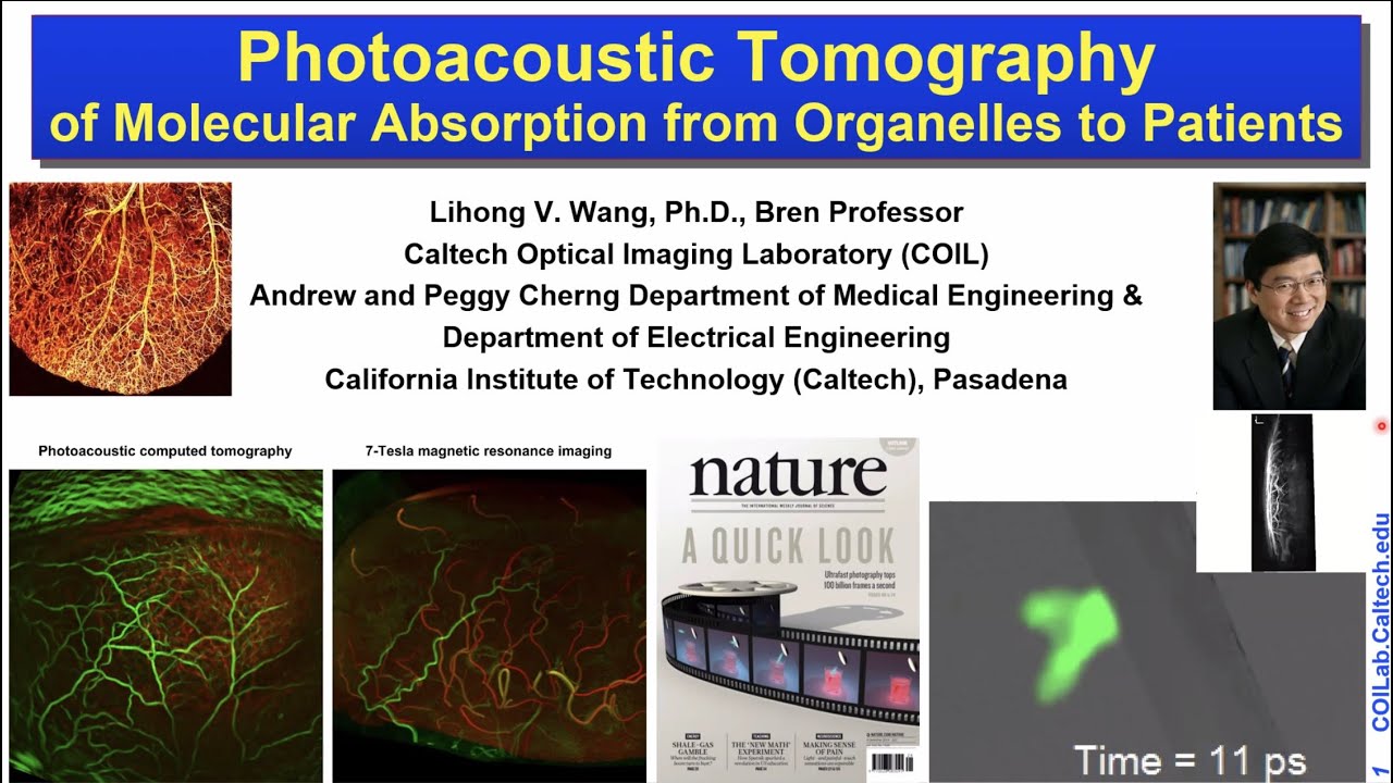
Photoacoustic tomography (PAT) has been developed for in-vivo
functional, metabolic, molecular, and histologic imaging by physically
combining optical and ultrasonic waves. Broad applications include earlycancer
detection and brain imaging. High-resolution pure optical imaging is
limited to superficial imaging within the optical diffusion limit (~1 mm in the
skin) in scattering tissue. By synergistically combining light and sound, PAT in
the form of either photoacoustic computed tomography or photoacoustic
microscopy breaks through this limit and provides deep penetration at high
ultrasonic resolution and high optical contrast. PAT is the only modality
capable of in vivo imaging across the length scales of organelles, cells, tissues,
and organs (or small-animal organisms) with consistent molecular contrast.
The US FDA has approved PAT in 2021 for breast cancer diagnosis. The
annual conference on PAT has become the largest in SPIE’s 20,000-attendee
Photonics West since 2010. In addition, compressed ultrafast photography, the
world’s fastest real-time camera, will be touched upon.