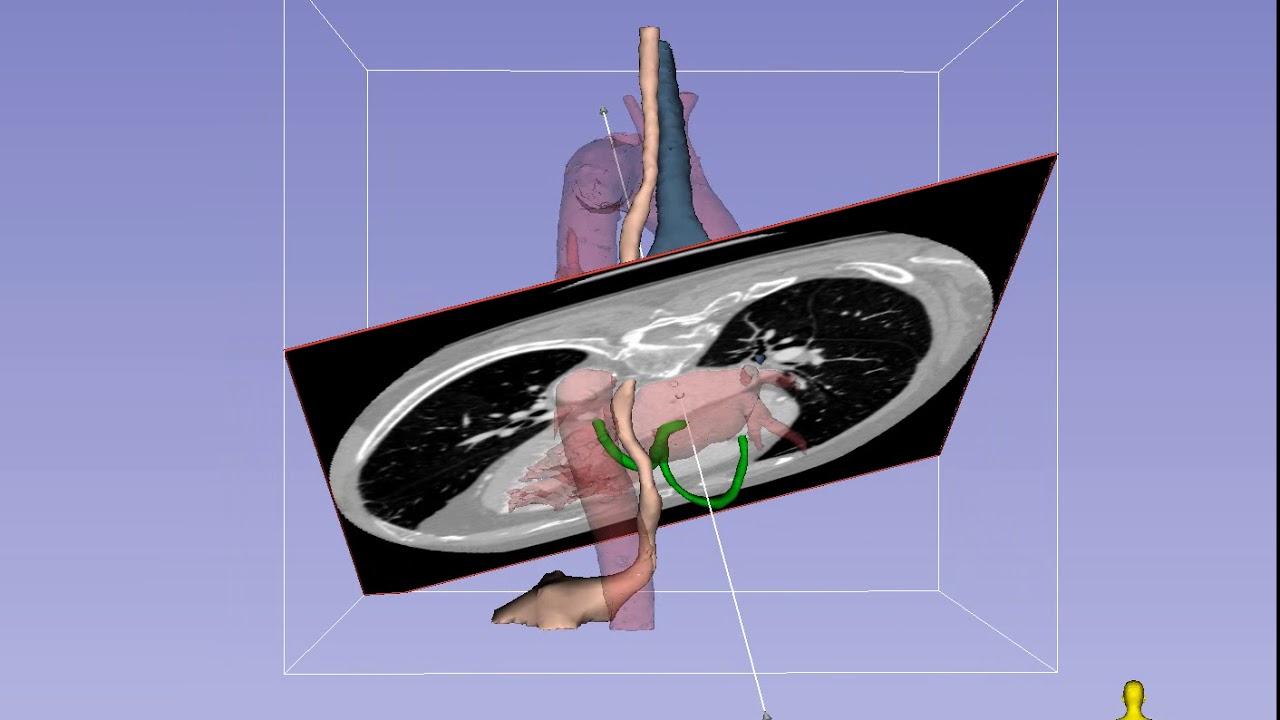
This series illustrates imaging planes corresponding to standard transesophageal echocardiography (TEE) views. In many cases the imaging planes also correspond to standard view in transthoracic echocardiography (TTE).
3D model based on cardiac-gated CT scan at 75% of R-R interval (mid-late diastole).
Segmentation and modeling in 3D Slicer ( [ Ссылка ] ).
= Legend = (not all structures are visible in all clips in the series)
Green : Heart Base (see [ Ссылка ] )
Dark red : Left heart and aortic blood pool
Pink : Upper GI tract including esophagus and stomach where the TEE probe resides
Dark blue : Tracheobronchial Tree (main source of TEE blind spots because of air contained therein)
Developed by Azad Mashari & Joshua Qua Hiansen at the Lynn and Arnold Irwin Advanced Perioperative Imaging Lab, Toronto General Hospital