Anatomy of the Urinary Bladder ❤️🩺
Location
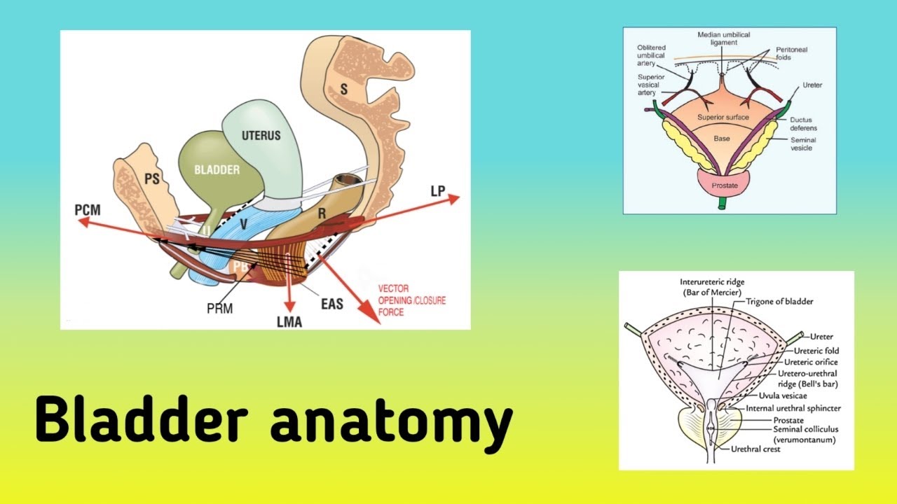
The urinary bladder is a hollow, muscular organ situated in the pelvis. In adults, it is located posterior to the pubic symphysis and anterior to the rectum in males and the vagina and uterus in females. When empty, the bladder is positioned entirely within the pelvis, but as it fills, it rises into the abdominal cavity.
Relations
In Males:
* Superior: Sigmoid colon and small intestine
* Anterior: Pubic symphysis
* Posterior: Rectum
* Inferior: Prostate gland (base of the bladder)
In Females:
* Superior: Uterus and small intestine
* Anterior: Pubic symphysis
* Posterior: Vagina and cervix
* Inferior: Urogenital diaphragm
Blood Supply ❤️🩸
The urinary bladder receives arterial blood from the following sources:
* Superior Vesical Arteries: Branches of the internal iliac arteries that supply the upper part of the bladder.
* Inferior Vesical Arteries (males): Branches of the internal iliac arteries that supply the lower part of the bladder, including the base and the prostatic urethra.
* Vaginal Arteries (females): Branches of the internal iliac arteries that supply the lower part of the bladder.
The venous drainage corresponds to the arterial supply:
* Vesical Venous Plexus: Drains into the internal iliac veins.
Nerve Supply 🧠
The bladder’s nerve supply is complex, involving both autonomic (sympathetic and parasympathetic) and somatic components.
Autonomic Nervous System:
* Parasympathetic Supply: Originates from the pelvic splanchnic nerves (S2-S4) and is responsible for bladder contraction (detrusor muscle) during urination (micturition).
* Sympathetic Supply: Originates from the hypogastric plexus (T11-L2) and is responsible for the relaxation of the bladder wall and contraction of the internal urethral sphincter during urine storage.
Somatic Nervous System:
* Pudendal Nerve: Provides somatic innervation to the external urethral sphincter, allowing voluntary control over urination.
Sensory Innervation:
* Sensory fibres from the bladder mucosa and detrusor muscle travel along the pelvic splanchnic nerves to provide feedback on bladder fullness and initiate the micturition reflex.
Summary ❤️🩺
The urinary bladder is a vital organ located in the pelvis, crucial for urine storage and voiding. Its anatomical relations differ slightly between males and females, primarily due to reproductive structures. The blood supply is chiefly derived from branches of the internal iliac arteries, with corresponding venous drainage. Nerve supply involves both autonomic and somatic systems, enabling involuntary and voluntary control over bladder function.
⏰ Chapter Timestamps:
00:00 - Introduction
00:30 - Location of the Bladder
03:05 - Shape and surfaces of the Bladder
06:20 - Relations
10:40 - Interior Aspect of the Bladder
13:05 - Blood Supply
14:02 - Nerve Supply
#UrinaryBladder
#Anatomy
#PelvicOrgans
#BladderLocation
#BladderRelations
#BloodSupply
#NerveSupply
#Urology
#MedicalAnatomy
#HumanBody
#OrganSystem
#UrogenitalSystem
#BladderFunction
#MedicalEducation
#Healthcare
#Physiology
#MedicalScience
#HumanAnatomy
#MedicalTerminology
#MedicalStudents