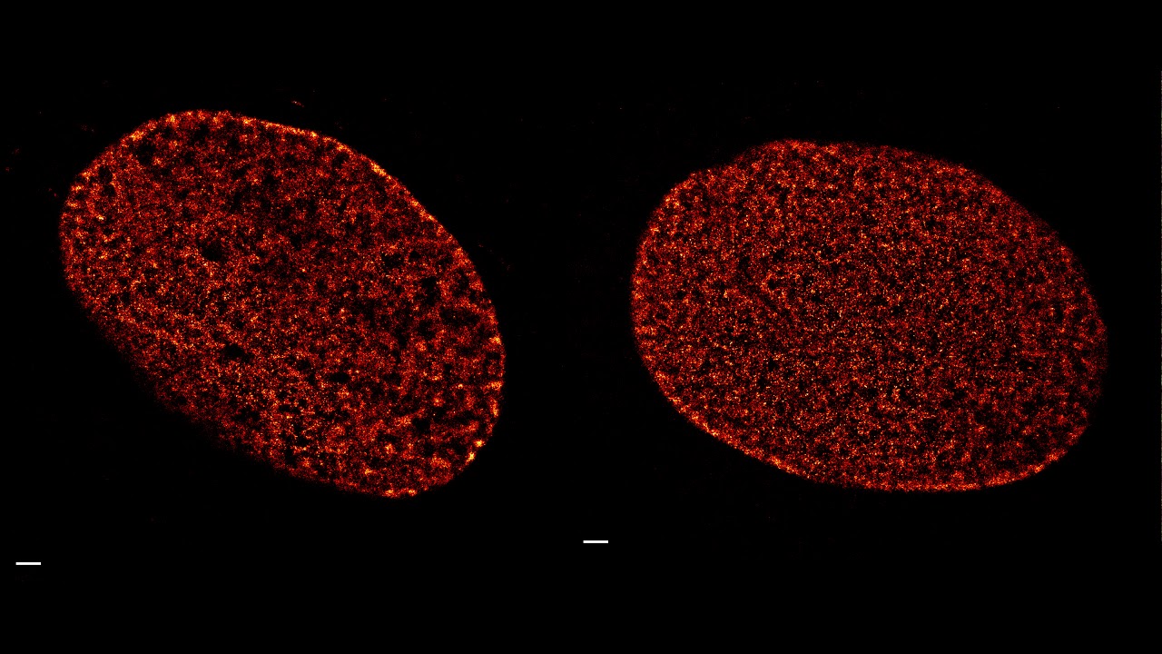
Two examples of TIRF single molecule localization microscopy (SMLM) time-lapse series of human fibroblast nucleus stained with 5-HMSiR-Hoechst. Living cells stained for 1h at 37°C in DMEM + 10% FBS and no washing of the probe applied before imaging. Each frame data set consisted of 10000 frames acquired at 100 Hz with excitation light intensity 18 kW/cm2.
Read more: [ Ссылка ]