Dr. Ebraheim’s educational animated video describes Intra - Articular fractures of the calcaneus and its classifications according to the Essex Lopresti.
Anatomy of the calcaneus
The calcaneus is the largest and most frequently fractured of the tarsal bones.
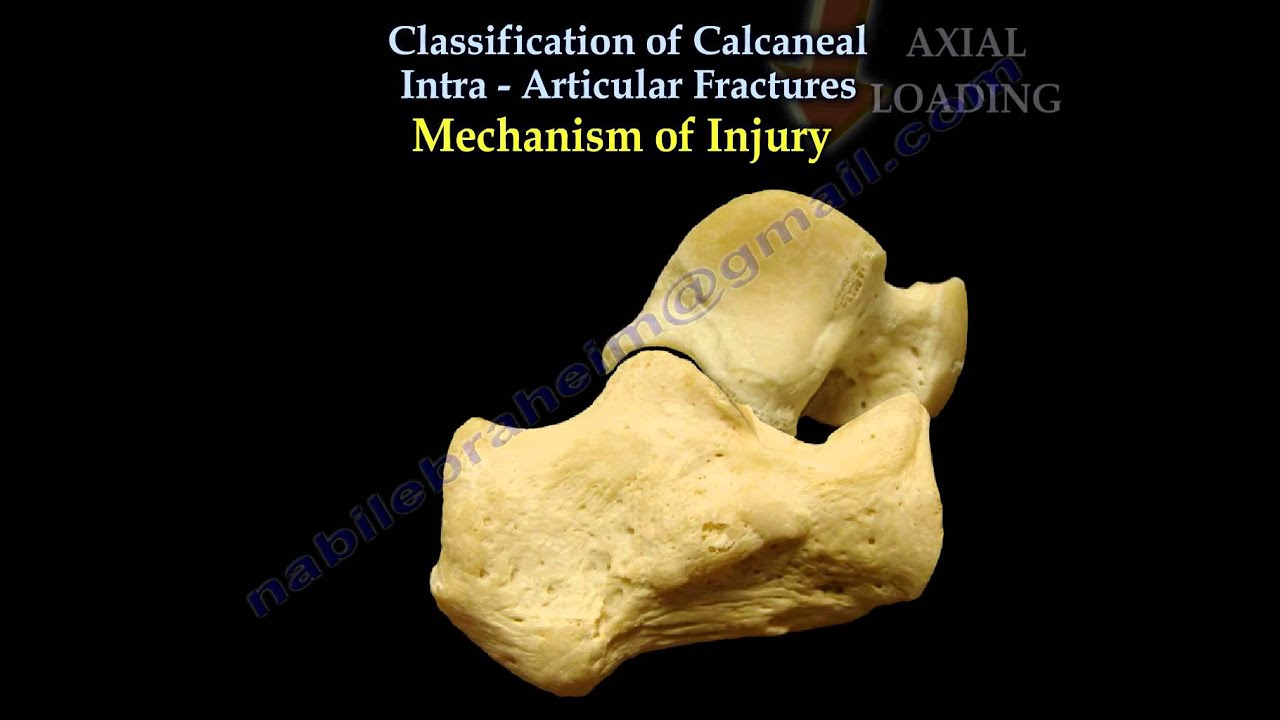
Mechanism of injury
•High energy injuries
•Usually due to fall
Results in axial loading
Lateral process of the talus acts as a wedge resulting in primary fracture line and constant medial fragment.
Lumbar spine injury 3-15%
Compartment syndrome 10%
Pathoanatomy
Heel in varus with height shortening and widening. With continued axial force, the tuberosity fragment rotates into varus.
Essex Lopresti classification
Divided into two types based on the location of the secondary fracture line which creates a free lateral piece of posterior facet separate from the tuberosity fragment.
•Joint depression: primary fracture line splits the calcaneus obliquely through the posterior facet and exits anterolaterally and posteromedially. Divides the calcaneus into anteromedial (sustentacular) and posterolateral (tuberosity fragments). Secondary fracture line exits superiorly behind the posterior facet. The posterior facet is a free fragment.
•Tongue type: primary fracture line exits anterolaterally and posteromedially. Secondary fracture line appears beneath the posterior facet and exits posteriorly through the tuberosity. The posterior facet is attached to the tuberosity.
In my opinion, the tongue type fracture can be treated with minimally invasive techniques. Tongue type fracture probably has a better outcome than a joint depression fracture.
Become a friend on facebook:
[ Ссылка ]
Follow me on twitter:
[ Ссылка ]