Extraocular Muscles, Movements Extrinsic Eye Muscles | 3D Human Anatomy | Organs
*****************************************************************
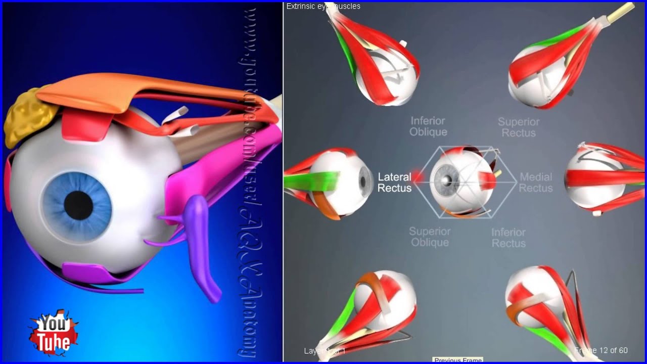
The extraocular muscles are the six muscles that control movement of the eye (there are four in bovines[citation needed]) and one muscle that controls eyelid elevation (levator palpebrae). The actions of the six muscles responsible for eye movement depend on the position of the eye at the time of muscle contraction.
Since only a small part of the eye called the fovea provides sharp vision, the eye must move to follow a target. Eye movements must be precise and fast. This is seen in scenarios like reading, where the reader must shift gaze constantly. Although under voluntary control, most eye movement is accomplished without conscious effort. Precisely how the integration between voluntary and involuntary control of the eye occurs is a subject of continuing research.[1] It is known, however, that the vestibulo-ocular reflex plays an important role in the involuntary movement of the eye.
Intermediate directions are controlled by simultaneous actions of multiple muscles. When one shifts the gaze horizontally, one eye will move laterally (toward the side) and the other will move medially (toward the midline). This may be neurally coordinated by the central nervous system, to make the eyes move together and almost involuntarily. This is a key factor in the study of strabismus, namely, the inability of the eyes to be directed to one point.
There are two main kinds of movement: conjugate movement (the eyes move in the same direction) and disjunctive (opposite directions). The former is typical when shifting gaze right or left, the latter is convergence of the two eyes on a near object. Disjunction can be performed voluntarily, but is usually triggered by the nearness of the target object. A "see-saw" movement, namely, one eye looking up and the other down, is possible, but not voluntarily; this effect is brought on by putting a prism in front of one eye, so the relevant image is apparently displaced. To avoid double vision from non-corresponding points, the eye with the prism must move up or down, following the image passing through the prism. Likewise conjugate torsion (rolling) on the anteroposterior axis (from the front to the back) can occur naturally, such as when one tips one's head to one shoulder; the torsion, in the opposite direction, keeps the image vertical.
The muscles show little inertia - a shutdown of one muscle is not due to checking of the antagonist, so the motion is not ballistic.
*****************************************************************
The description of: [ Ссылка ]
Our video review of human anatomy: [ Ссылка ]
*****************************************************************
Human anatomy (gr. ἀνατομία, "dissection", from ἀνά, "up", and τέμνειν, "cut") is primarily the scientific study of the morphology of the human body. Anatomy is subdivided into gross anatomy and microscopic anatomy (histology) Gross anatomy (also called topographical anatomy, regional anatomy, or anthropotomy) is the study of anatomical structures that can be seen by the naked eye. Microscopic anatomy involves the use of microscopes to study minute anatomical structures, and is the field of histology which studies the organization of tissues at all levels, from cell biology (previously called cytology), to organs. Anatomy, human physiology (the study of function), and biochemistry (the study of the chemistry of living structures) are complementary basic medical sciences, that are generally taught together (or in tandem) to students studying medicine.